Demystifying Medical Imaging Technology

Medical imaging diagnostics is a specialized field of medicine. It uses a variety of advanced technical means to form an image of the human body, allowing doctors the information they need to make accurate diagnoses and treatment plans. I believe that many people who have visited a hospital have had some experience in taking an x-ray or ultrasound examination. Others may have heard of computed tomography (CT), Magnetic Resonance Imaging (MRI), and other terms, but are not really familiar or clear on the principal differences between these detection methods. Often questions arise such as, “Will taking a CT or MRI make me feel sick? Will the radiation exposure cause harm to me or my child?” In this article, I will briefly introduce Beijing United Family Hospital’s most commonly used medical imaging procedures: x-ray, MRI, CT, and ultrasound. Hopefully this will help answer some of the most common questions and alleviate some of the fear and uncertainty surrounding radiology.
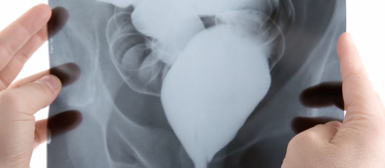
X-Ray
In 1895 while researching electrical charges in various types of vacuum tubes, a German scientist named Wilhelm Conrad Röntgen discovered “a new kind of ray.” He temporarily named in an “X-ray” as it was still as yet unknown. The name stuck, and within just a few months time, x-rays were starting to be applied to medical imaging. In 1896, the Royal Hospital in Glasgow, Scotland established the world’s first Radiology Department.
Today, an x-ray is one of the most common imaging methods used by hospitals throughout the world. The image is produced when radiation is directed at parts of the human body, with these x-rays being absorbed by different tissues and bones in different amounts, producing black and white pictures showing what lies underneath our skin. X-rays can be used to diagnose a variety of bone issues, such as fractures, and lung diseases such as pneumonia, lung cancer or emphysema. Abdominal x-rays can help detect intestinal obstruction, gasses within the body cavity, and certain types of kidney stones.
The radiation dose of a chest x-ray image is about 0.15 mSv. (Note that “mSV” means millisievert, or 0.001 Sieverts, which is a measure of radiation named after a Swedish Scientist. This means a typical chest x-ray exam gives a dose of about 0.00015 Sieverts). In our daily lives, we are affected by cosmic radiation in typical doses of about 2 mSv per year. In[1] routine medical use, x-ray radiation dosage is carefully controlled to be highly secure and safe. With the latest advances in technology, today’s X-ray machines are ever safer and more effective, while also being fast and inexpensive for patients. This makes it a widespread choice for doctors to quickly and easily help with diagnoses, earning the x-ray a unique position in medical imaging.
CT
Computed tomography imaging or “CT,” is a digital reconstruction of multiple radiation images. It is produced when different groups of radiation waves are beamed into the body, creating several levels of images as a result of the different levels of bodily absorption. These images are then reconstructed digitally with the aid of a computer, and presented in a layered manner, providing a reconstructed cross section of structures within the human body. By digitally adding multiple layers into a cross section, radiology technicians and doctors can effectively examine a three-dimensional image of the problem area, noting the specific characteristics and nuances from every angle. This high-resolution technology is incredibly convenient for doctors in making diagnoses.
Images from CT scans can be used to diagnose tumors, inflammation, and pain in all parts of the body. Internal organs, bones, soft tissues, and blood vessels all appear clearer in a CT image than in x-ray images. Therefore, CT scans can have particular advantages when doctors are seeking to understand the effects of complex problems such as a brain hemorrhage, pulmonary and cardiovascular disease, a complex bone fracture, or kidney and gall stones.
MRI
Approximately 70% of the human body is composed of water. MRI (Magnetic Resonance Imaging) technology relies on the stimulation of protons in water. When the body, with its majority liquid makeup, is placed in a magnetic field with the appropriate electromagnetic wave exposure, it forces hydrogen protons to resonate and change their orientation. This allows for the release of electromagnetic waves that can be analyzed. Because different groups of protons can produce different electromagnetic signals, when processed by computers, doctors can gain a unique picture of internal body structures.
An MRI scan can produce an image with excellent resolution of soft tissues. It is also useful to doctors in looking at solid organs, such as cranial nerve systems, musculoskeletal systems, spinal cartilage, and thyroid glands. It has excellent diagnostic value when looking at the liver, bile, spleen, kidneys, pancreas, adrenal glands, and reproductive organs (such as the bladder, uterus, ovary, and prostate, as well as breasts). Compared with a CT scan, MRI offers safer imaging without radiation, but a longer scan time is required as the patient lies within the MRI machine for several minutes. Since MRI systems produce a strong magnetic field, patients undergoing MRI exams are required to remove all metal jewelry. An MRI exam is not suitable for some patients who have metal implants within their body, such as those with pacemakers, or certain hip replacements.
Ultrasound
High frequency sound waves are used in medical ultrasound imaging, whereby sound is projected at human tissue and echo signals bounces back, forming a group of signals that computers can generate into a comprehensive image. Ultrasound exams are useful in examining solid organs of the human body, such as the liver, gallbladder, pancreas, spleen, kidneys, and uterus. Ultrasound is also useful for getting a look at organs like the thyroid gland, mammary glands, eyes, and cardiovascular system. It helps with the detection of lesions under the skin, and is often used for fetal and prenatal examinations.
Due to the convenient non-invasive and non-radiation nature of the ultrasound, it is widely used in hospitals. The ultrasound, however, is not useful in diagnosing issues with bones or gas-bearing organs such as the lungs.
When administered properly, the benefits of imaging exams normally far outweigh the possible risks to patients, depending on their situation. These above four kinds of medical imaging normally do not cause pain. For certain CT and MRI examinations, patients are sometimes asked to take substances that can help compare and contrast different organs and tissues, allowing for a better understanding and clearer pictures for doctors. In these cases, patients might be asked to take the substances orally, by intravenous drip, or through an enema, which may cause temporary discomfort. As well, a small number of patients may find it difficult to remain still during the examination process, such as those with chronic pain, acute claustrophobia, or other such discomfort. For these patients and under a doctor’s direct supervision, pain-killing or other pharmaceuticals may be administered to help stabilize them so that the examination can be completed. Female patients that are pregnant should always inform the radiologist before scheduling an exam.
If you or your child has already previously completed an imaging examination, please inform your radiologist so that they can check the results and avoid repeating the exam if not necessary. We suggest you communicate fully with the radiologist or technician all your past histories and any special situations before the examination. This will allow radiology staff to be more attuned to your wishes, provide you with more details, and generally tell you about which things you may need to be aware.
[1] According to the International Commission on Radiological Protection, 1 Sievert carries with it a 5.5% chance of eventually developing cancer, so a chest x-ray would have a very small percentage chance of 0. 000825% or so to possibly cause harm.
FOLLOW US HERE:
 中文微信
中文微信  BJU English WeChat
  微商城WeChat Store
  微博
微博 Copyright United Family Healthcare 2014 All right reserved - 京卫网审[2014]第1927号 - 京ICP备13017554号-4